 English
 Français
 Español
 Deutsch
 Italiano
 العربية



هاتف
:+86-755-86961139
بريد إلكتروني
:sales@vivo-light.com
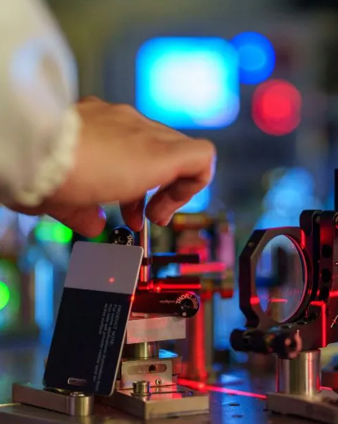

Establishing peripheral intravenous access in geriatric patients within psychiatric inpatient units presents significant operational and procedural challenges. Age-associated vascular deterioration, chronic pharmacotherapy, cognitive impairment, and involuntary physical agitation frequently lead to Difficult Intravenous Access (DIVA). In these clinical scenarios, multiple cannulation attempts not only increase physical trauma and distress for vulnerable patients but also prolong bedside nursing procedures and elevate the risk of extravasation or phlebitis.
To address these clinical bottlenecks, projection-type near-infrared (NIR) vein illumination has become an established, non-invasive procedural adjunct. By projecting a real-time, in situ map of superficial vasculature directly onto the patient's skin, this optical technology assists clinical staff in locating veins, evaluating vessel course, and identifying bifurcations prior to needle insertion.
This article examines published clinical evidence evaluating the application of projection-type NIR vascular imaging in elderly psychiatric inpatients undergoing peripheral intravenous indwelling catheterization.
Venipuncture is among the most common invasive procedures performed by nursing staff. However, elderly psychiatric patients present specific pathophysiological and psychological characteristics that complicate vascular access:
1. Vascular Sclerosis and Fragility: Aging blood vessels lose elasticity, develop hardened or thickened walls, and feature diminished surrounding subcutaneous connective tissue. These veins tend to roll easily upon needle contact, collapse readily, or rupture under mechanical pressure.
2. Behavioral Agitation and Limited Cooperation: Patients diagnosed with conditions such as senile dementia, schizophrenia, mood disorders, or organic mental disorders often struggle to follow procedural commands. Fear, perceptual distortion, or motor restlessness can lead to sudden limb withdrawal during cannulation.
3. Compromised Peripheral Vasculature from Chronic Regimens: Long-term administration of psychotropic medications, frequent diagnostic blood draws, and repeated infusion therapies often result in progressive vascular fibrosis, leaving few palpable or visible superficial veins available.
Under conventional manual palpation protocols, nurses rely on tactile perception and surface landmarks. In elderly psychiatric patients with deep, non-palpable, or highly mobile veins, traditional blind cannulation often requires repeated attempts, which can escalate patient agitation and delay timely medical interventions.
A comparative clinical study published in the peer-reviewed nursing journal Today Nurse systematically assessed the clinical utility of projection-type near-infrared vascular imaging compared with conventional palpation methods.
• Clinical Setting & Study Cohort: The study was conducted in an inpatient psychiatric department between February 2018 and February 2019, involving 63 elderly patients with mental disorders requiring multi-day intravenous therapy.
• Inclusion Criteria: Patients aged ≥ 60 years meeting ICD-10 diagnostic criteria for mental disorders, anticipated intravenous infusion duration exceeding 5 days, requiring a 24G closed peripheral intravenous indwelling needle (BD Intima-II), and presenting vascular condition assessment scores ≥ 5 points (evaluated across vein elasticity, visibility, fullness, fixation, and mobility).
• Specific Device Investigated: Vivolight Projection-Type Near-Infrared Vascular Imaging System developed by Shenzhen Vivolight Medical Device Co., Ltd.
• Control Group (February 2018 – July 2018): 31 patients (totaling 68 venipuncture procedures) managed using conventional anatomical visualization and manual palpation.
• Observation Group (August 2018 – February 2019): 32 patients (totaling 72 venipuncture procedures) managed with the assistance of the Vivolight projection vascular imaging system.
Baseline demographic and clinical parameters—including age, gender distribution, disease classification, and initial vascular condition scores—showed no statistically significant differences between the two cohorts (P > 0.05), ensuring baseline clinical comparability.
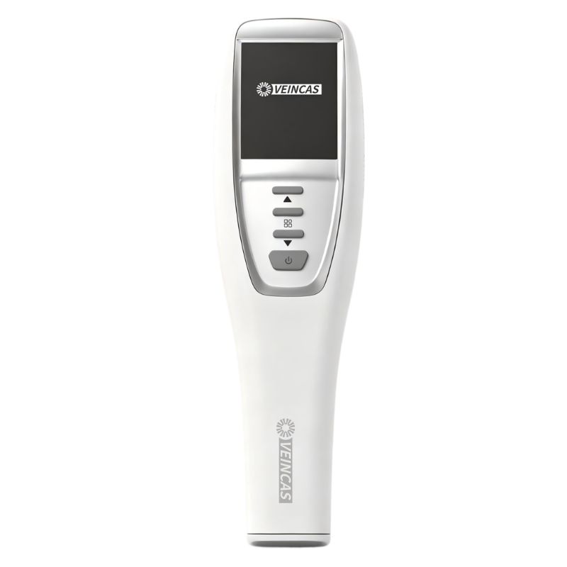
Projection vein finder for clinical vascular visualization
The Vivolight projection vein finder relies on the differential optical absorption of near-infrared light by physiological tissues:
• Deoxygenated hemoglobin circulating within subcutaneous venous blood possesses a significantly higher absorption coefficient for near-infrared light wavelengths than surrounding biological tissues, such as skin, fat, and fascia.
• The system's optical sensor detects the reflected near-infrared signal differences, processes the vascular contrast map via dedicated image algorithms, and projects an accurate, real-time image directly back onto the cutaneous surface in a precise 1:1 anatomical scale.
• This allows healthcare providers to assess vessel trajectory, track bifurcations, evaluate localized depth variations, and avoid venous valves before initiating puncture.
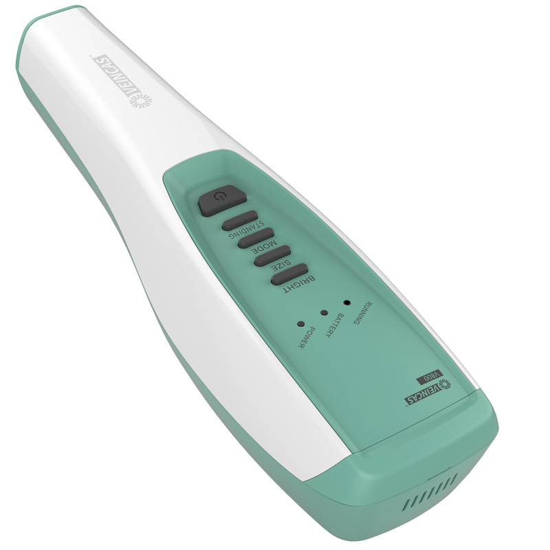
Projection vein finder device
The study analyzed three core clinical indicators: first-attempt venipuncture success rate, total puncture procedure duration, and overall patient/family satisfaction.
| Clinical Metric | Control Group (Manual) | Observation Group | Test Statistic | P-value |
|---|---|---|---|---|
| First-Attempt Success Rate | 70.59% (48/68) | 84.72% (61/72) | χ² = 4.052 | P = 0.044 (P < 0.05) |
| Puncture Procedure Time | 4.74 ± 0.84 min | 4.24 ± 1.08 min | t = 3.045 | P = 0.003 (P < 0.01) |
| Patient & Family Satisfaction | 74.19% (23/31) | 93.75% (30/32) | χ² = 4.510 | P = 0.034 (P < 0.05) |
The statistical findings highlight three key operational improvements:
1. Higher First-Attempt Success Rate: Successful first-attempt cannulation occurred in 84.72% of procedures using the projection vein finder, compared to 70.59% with manual palpation (P = 0.044). This represents a statistically significant 14.13 percentage point increase in initial puncture success among clinically difficult veins.
2. Reduction in Bedside Procedure Time: Procedure time was reduced from 4.74 ± 0.84 minutes in the control group to 4.24 ± 1.08 minutes in the observation group (P = 0.003). In psychiatric nursing, a 30-second reduction meaningfully decreases the duration of physical restraint or limb immobilization required during needle advancement.
3. Enhanced Inpatient and Family Satisfaction: Total satisfaction reached 93.75% in the projection vein finder cohort compared to 74.19% in the manual cohort (P = 0.034), driven by fewer repeat needle passes and reduced psychological anxiety during cannulation.
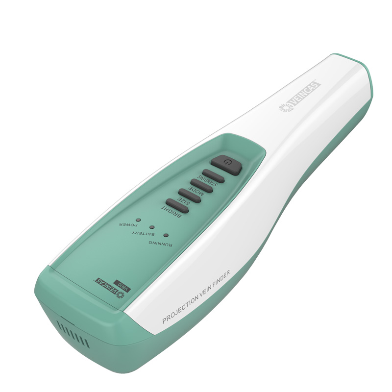
Explore the Vivolight product range.
While near-infrared projection technology provides clear operational advantages, clinical evidence supports several practical considerations:
1. Procedural Adjunct, Not a Mechanical Substitute: Near-infrared projection aids in surface localization and mapping of superficial venous geometry. It does not eliminate the need for clinical palpation to verify vein elasticity, compressibility, or depth.
2. Tissue Depth and Anatomical Limitations: Near-infrared light penetration is limited by physics to superficial subcutaneous layers (typically within 8–10 mm). In clinical cases marked by severe localized edema, thick subcutaneous adipose tissue, or organized hematomas, optical contrast may be attenuated, requiring complementary assessment or ultrasound guidance.
3. Ergonomic Hands-Free Application: The projection vein finder mobile stand configuration allows non-contact, hands-free vascular projection. This design enables nurses to dedicate both hands to limb stabilization, vein traction, and aseptic catheter insertion, which is critical when managing restless or uncooperative patients.
[1] Gu, L. Q. (2020). Application of projection-type infrared vascular imaging device in peripheral venous indwelling needle puncture for elderly patients with mental disorders Today Nurse, 27(36), 153–155. DOI: 10.19793/j.cnki.1006-6411.2020.36.062.
[2] Infusion Nurses Society (INS). (2021). Infusion Therapy Standards of Practice (8th Edition). Journal of Infusion Nursing, 44(1S), S1–S224.
[3] Zhang, Y., & Liu, H. (2018). Clinical evaluation of infrared vascular imaging technology in difficult peripheral intravenous access: A comparative study. Chinese Journal of Modern Nursing, 24(4), 80–81.
[4] Schults, J., et al. (2019). Technologies to facilitate peripheral intravenous catheter insertion in difficult access patients: A systematic review and meta-analysis. International Journal of Nursing Studies, 98, 108–117.
1. What is a projection-type vein finder?
A projection-type vein finder uses near-infrared imaging to visualize superficial veins and project a real-time vein map onto the skin, helping healthcare professionals assess potential venipuncture sites.
2. How can a vein finder support venipuncture in elderly patients?
It can help clinicians identify superficial vein pathways when veins are difficult to see, potentially reducing repeated puncture attempts. Device use should be combined with clinical assessment and established venipuncture protocols.
3. Can near-infrared vein imaging replace palpation?
No. Vein imaging is an adjunct to clinical assessment. Palpation remains important for evaluating characteristics such as vein elasticity, compressibility, and depth.
4. What limitations should clinicians consider when using a vein finder?
Near-infrared imaging primarily visualizes superficial vascular structures. Edema, thick subcutaneous tissue, and other anatomical factors may affect image quality, so additional assessment or ultrasound guidance may be appropriate in selected cases.
5. Where can healthcare providers learn more about projection vein finders?
Healthcare providers can review the projection vein finder range and individual device specifications on the Vivolight official website.
ترك رسالة
مسح ضوئي إلى WhatsApp :


 IPv6 الشبكة المدعومة