 English
 Français
 Español
 Deutsch
 Italiano
 العربية




تطبيق تبادل بيانات التصوير المقطعي البصري في الممارسة السريرية الحقيقية في مستشفى بكين فواي
لطالما ازدهر طب القلب التداخلي بفضل التبادل المعرفي، متجاوزًا الحدود ومستويات الخبرة والثقافات السريرية. وفي مؤتمر CIT 2026، أحد أهم مؤتمرات طب القلب التداخلي في منطقة آسيا والمحيط الهادئ، كان هذا التبادل جليًا في كل جلسة، وكل نقاش، وكل حديث جانبي. لكن بالنسبة لمجموعة من الأطباء الدوليين، لم يكن الجزء الأهم من زيارتهم داخل قاعة المؤتمر، بل داخل مختبر القسطرة.
بعد برنامج مؤتمر CIT 2026، شركة فيفولايت الطبية سافر وفد دولي من الأطباء إلى مستشفى فواي التابع للأكاديمية الصينية للعلوم الطبية، وهو المركز الرائد في الصين لأمراض القلب والأوعية الدموية، للمشاركة في برنامج تبادل سريري داخل المستشفى يركز على التدخل التاجي عن طريق الجلد الموجه بالتصوير داخل الأوعية الدموية. بالنسبة لهؤلاء الأطباء الزائرين، مثّلت فرصة التعرّف على أحد أكثر مختبرات القسطرة استخدامًا في العالم، وملاحظة كيفية دمج التصوير في الممارسة اليومية، أمرًا لا يمكن لأي محاضرة أو ندوة أن تحاكيه: فرصة لرؤية، في الوقت الفعلي، كيف تُترجم التكنولوجيا إلى قرارات سريرية تؤثر على نتائج المرضى.
داخل مختبر القسطرة: مراقبة التدخل التاجي عن طريق الجلد الموجه بالتصوير في الوقت الحقيقي
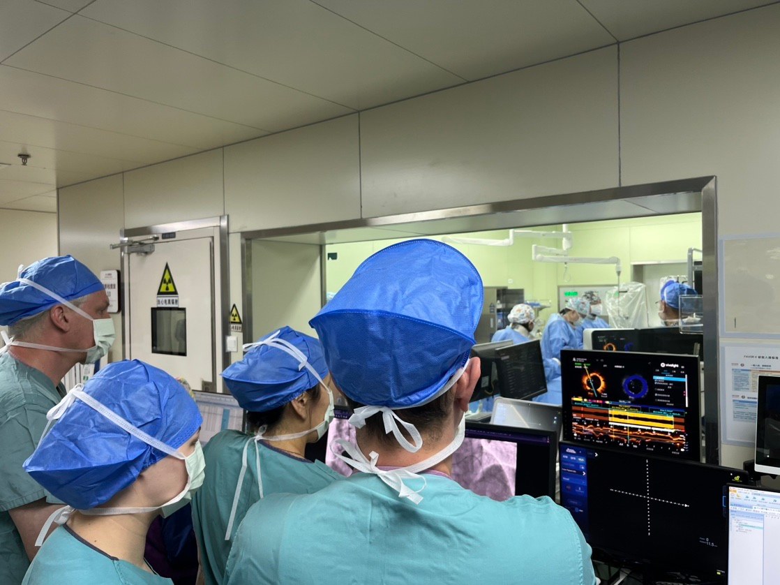
في 22 أبريل، بدأ التبادل السريري في المكان الأكثر أهمية - غرفة التحكم في مختبر القسطرة. شاهد الوفد إجراءً مباشرًا أجراه البروفيسور سونغ لي، متابعًا سير العمل السريري عن كثب. منذ لحظة إدخال قسطرة التصوير المقطعي التوافقي البصري (OCT) وحتى الحصول على الصور وتفسيرها في الوقت الفعلي، اكتسب المشاركون رؤية مباشرة لكيفية دعم معلومات التصوير داخل الأوعية الدموية لفهم الإجراءات في بيئة تدخلية حقيقية.
.
بالنسبة للكثيرين في الوفد، كانت هذه فرصة نادرة لمراقبة كيفية دمج المشغل الخبير لبيانات التصوير في كل خطوة من خطوات الإجراء - ليس كفكرة لاحقة أو أداة لحل المشكلات، ولكن كجزء أساسي من سير العمل السريري.
من الملاحظة إلى الحوار: مناقشة قائمة على دراسة حالة
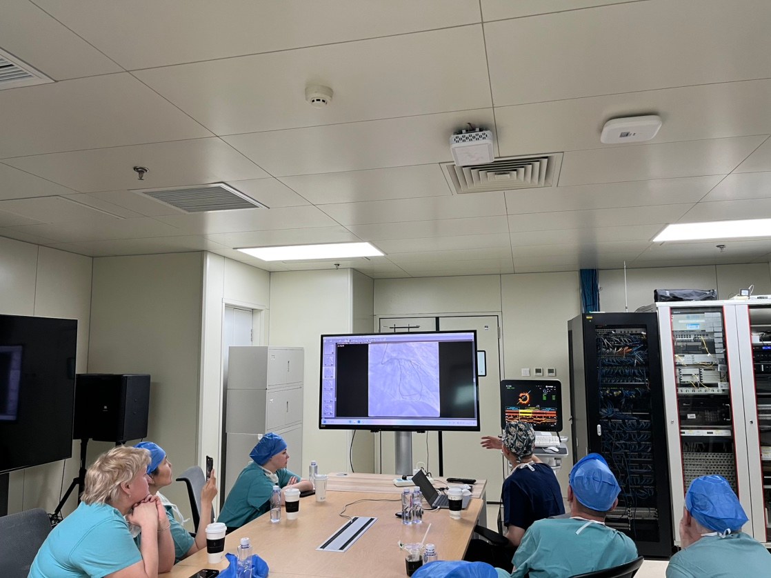
بعد الإجراء، انضم الوفد إلى البروفيسور سونغ في جلسة نقاش معمقة تمحورت حول حالات رأب الأوعية التاجية عن طريق الجلد (PCI) الواقعية. لم تكن محاضرة، بل حوارًا تفاعليًا، حيث طرح الأطباء الزائرون وجهات نظرهم السريرية، وطرحوا أسئلة مستمدة من ممارساتهم اليومية، وانخرطوا في تبادل صريح قائم على دراسة الحالات، وهو ما لا يحدث إلا عندما يجلس أطباء من خلفيات مختلفة معًا أمام بيانات تصوير حقيقية على الشاشة.
شمل الحوار سيناريوهات ذات صلة سريرية، بما في ذلك:
● تضيق الشريان داخل الدعاماتكيف يمكن لتقنية التصوير المقطعي البصري (OCT) أن توفر تصويرًا عالي الدقة للنتائج المتعلقة بالدعامات والأنسجة، مما يساعد الأطباء على فهم الآليات المحتملة وراء إعادة التضيق بشكل أفضل - سواء كان ذلك فرط تنسج البطانة الداخلية، أو عدم تمدد الدعامات، أو كسر الدعامات - وتصميم العلاج وفقًا لذلك.
● تكلس شديد غير مركزيكيف يمكن أن يساعد التصوير المقطعي البصري في تقييم توزيع الكالسيوم وسمكه وشكله وشدته، مما يوفر معلومات بالغة الأهمية لاختيار الاستراتيجية - من استئصال العصيدة الدوراني إلى تفتيت الحصى داخل الأوعية الدموية - وتحسين العلاج.
● تشريح حافة الدعامةكيف يمكن للتصوير عالي الدقة تحديد التسلخات والأورام الدموية التي قد لا تظهر في تصوير الأوعية الدموية، مما يمكّن الأطباء من اتخاذ قرارات مستنيرة بشأن ما إذا كان ينبغي العلاج أو التمديد أو المراقبة.
Tمن خلال مراجعة الحالات وحوار الخبراء، ناقش الفريق كيف يمكن للتصوير داخل الأوعية الدموية - وخاصة التصوير المقطعي التوافقي البصري (OCT) - أن يدعم سير عمل التدخل التاجي عن طريق الجلد (PCI) بالكامل: تقييم الآفة، والتخطيط الإجرائي، وتحسين العلاج، والتقييم بعد التدخل. وقد عززت المناقشة نقطةً لاقت صدىً واسعاً في مختلف الثقافات وبيئات الممارسة: التصوير لا يساعدك فقط على الرؤية بشكل أفضل، بل يساعدك أيضاً على اتخاذ قرارات أفضل.
منصة التصوير المقطعي البصري متعدد الوسائط P80: سحبة واحدة، إجابات متعددة
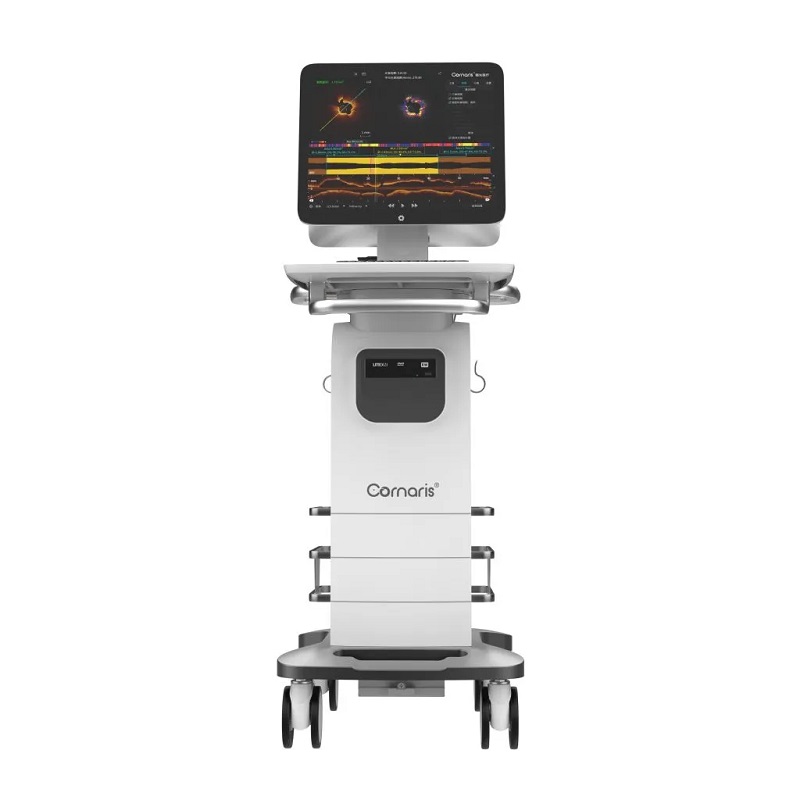
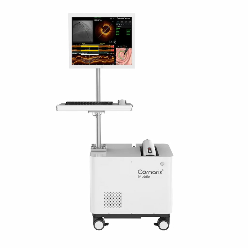
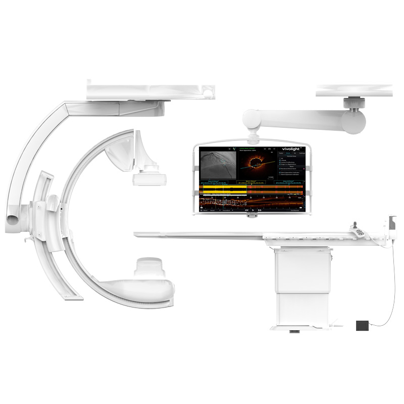
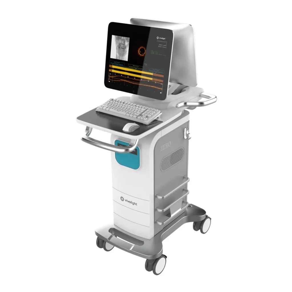
كما ربط التبادل السريري مناقشة الحالات الواقعية بشركة فيفولايت نظام التصوير المقطعي البصري متعدد الأنماط كورناريس P80. يوفر النظام حلاً شاملاً لفحص عنق الرحم يجمع بين التقييم الوظيفي وتقييم التصوير المقطعي التوافقي البصري من خلال عملية سحب واحدة للتصوير المقطعي التوافقي البصري:
● FFR المشتق من التصوير المقطعي التوافقي البصري: يجمع OCT-FFR بين التقييم الوظيفي المستمد مباشرة من بيانات OCT وتقييم OCT، مما يلغي الحاجة إلى سلك ضغط منفصل ويوفر معلومات ديناميكية الدم ضمن نفس سير عمل التصوير.
● مؤشر توهين اللويحة (IPA): تقييم استقرار اللويحات الذي يساعد الأطباء على تحديد خصائص تكوين اللويحات وتحديد الآفات المعرضة للخطر.
● التقييم الذكي للكالسيوم (ICA): تحديد حجم الكالسيوم ودرجة التكلس باستخدام الذكاء الاصطناعي، مما يدعم اختيار استراتيجية المعالجة المسبقة الدقيقة للآفات المتكلسة.
بفضل نتائج التقييم متعدد الوسائط المتاحة من خلال سحب واحد، تم تصميم منصة P80 لمساعدة الأطباء في الحصول على معلومات أكثر قابلية للتنفيذ دون مواد استهلاكية إضافية أو وقت إجراء إضافي - وهي ميزة عملية حظيت بتقدير خاص من قبل الوفد، حيث يعمل الكثير منهم في بيئات سريرية حيث تعتبر كفاءة الإجراءات أولوية يومية.
نظرة مستقبلية: من التبادل إلى الشراكة
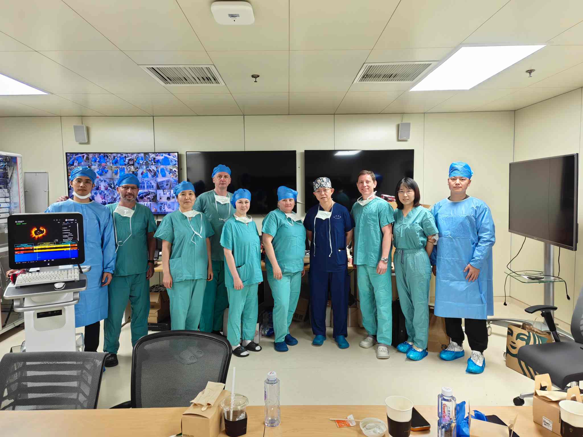
مع نهاية الزيارة، تجاوز النقاش الجوانب التقنية السريرية. ناقش الوفد وفريق البروفيسور سونغ إمكانية إقامة تعاون مستمر، يشمل تبادل الحالات في المستقبل، والأنشطة الأكاديمية المشتركة، ومواصلة الحوار حول التدخل التاجي عن طريق الجلد الموجه بالتصوير. كانت القيمة واضحة لكلا الطرفين: فاختلاف فئات المرضى، وأنماط الممارسة، وأنظمة الرعاية الصحية، كلها عوامل تُسهم في إثراء فهمنا للتحديات السريرية المشتركة. كان هذا التبادل في فواي بمثابة نقطة انطلاق، لا خاتمة.
من خلال الجمع بين التدريب الأكاديمي في المؤتمرات والتبادل السريري في المستشفيات، ساعدت الزيارة الوفد على بناء فهم أعمق لدور التصوير المقطعي التوافقي البصري (OCT) في جميع مراحل عملية التدخل التاجي عن طريق الجلد (PCI)، بدءًا من تقييم الآفة واختيار الاستراتيجية وصولًا إلى تحسين العلاج وتقييم ما بعد التدخل. ستواصل شركة Vivolight Medical العمل عن كثب مع الخبراء السريريين والمستشفيات والشركاء العالميين لتعزيز التعليم العملي والتواصل السريري والتطبيق الأوسع للتدخل التاجي الموجه بالتصوير المقطعي التوافقي البصري.
【تنصل】
إن محتوى هذه المقالة مخصص فقط للتبادل الأكاديمي وتبادل المعلومات على مستوى الصناعة ولا يشكل أي إعلان أو توصية بالمنتج أو نصيحة طبية.
ترك رسالة
مسح ضوئي إلى WhatsApp :

 IPv6 الشبكة المدعومة